

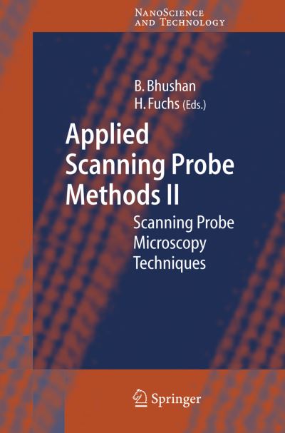
The Nobel Prize of 1986 on Sc- ningTunnelingMicroscopysignaled a new era in imaging. The sc- ning probes emerged as a new - strument for imaging with a p- cision suf?cient to delineate single atoms. At ?rst there were two - the Scanning Tunneling Microscope, or STM, and the Atomic Force Mic- scope, or AFM.
The STM relies on electrons tunneling between tip and sample whereas the AFM depends on the force acting on the tip when it was placed near the sample. These were quickly followed by the M- netic Force Microscope, MFM, and the Electrostatic Force Microscope, EFM. The MFM will image a single magnetic bit with features as small as 10nm.
With the EFM one can monitor the charge of a single electron. Prof. Paul Hansma at Santa Barbara opened the door even wider when he was able to image biological objects in aqueous environments.
At this point the sluice gates were opened and a multitude of different instruments appeared. There are signi?cant differences between the Scanning Probe Microscopes or SPM, and others such as the Scanning Electron Microscope or SEM. The probe microscopes do not require preparation of the sample and they operate in ambient atmosphere, whereas, the SEM must operate in a vacuum environment and the sample must be cross-sectioned to expose the proper surface.
However, the SEM can record 3D image and movies, features that are not available with the scanning probes.
| ISBN: | 9783540262428 |
| Publication date: | 21st February 2006 |
| Author: | Bharat Bhushan, Harald Fuchs |
| Publisher: | Springer an imprint of Springer Berlin Heidelberg |
| Format: | Hardback |
| Pagination: | 420 pages |
| Series: | Nanoscience and Technology |
| Genres: |
Electronics engineering Engineering applications of polymers and composites Condensed matter physics (liquid state and solid state physics) Spectrum analysis, spectrochemistry, mass spectrometry Nanotechnology Materials science |
The Nobel Prize of 1986 on Sc- ningTunnelingMicroscopysignaled a new era in imaging. The sc- ning probes emerged as a new - strument for imaging with a p- cision suf?cient to delineate single atoms.
Applied Scanning Probe Methods features in the following genres: Electronics engineering, Engineering applications of polymers and composites, Condensed matter physics (liquid state and solid state physics), Spectrum analysis, spectrochemistry, mass spectrometry, Nanotechnology, Materials science
Hardback. Not Available.
Applied Scanning Probe Methods was written by Bharat Bhushan, Harald Fuchs and published by Springer an imprint of Springer Berlin Heidelberg
Applied Scanning Probe Methods has 420 pages
Yes it is part of Nanoscience and Technology series